
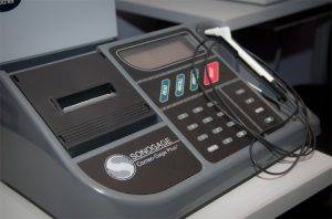
 Corneal Pachymetry
Corneal pachymetry is the process of measuring the thickness of the cornea. It is used to perform corneal pachymetry prior to refractive surgery, for Keratoconus screening, LRI surgery and is useful in screening for patients suspected of developing glaucoma among other uses.
Digital Retinal Imaging
We use cutting-edge digital imaging technology to assess your eyes. Many eye diseases, if detected at an early stage, can be treated successfully without total loss of vision.
Digital Retinal Imaging & OCT Scans
We use cutting-edge digital imaging technology to assess your eyes. Many eye diseases, if detected at an early stage, can be treated successfully without total loss of vision. Your retinal Images will be stored electronically. This gives the eye doctor a permanent record of the condition and state of your retina.
This is very important in assisting your Optometrist to detect and measure any changes to your retina each time you get your eyes examined, as many eye conditions, such as glaucoma, diabetic retinopathy and macular degeneration are diagnosed by detecting changes over time.
The advantages of digital imaging include:
- Quick, safe, non-invasive and painless
- Provides detailed images of your retina and sub-surface of your eyes
- Provides instant, direct imaging of the form and structure of eye tissue
- Image resolution is extremely high quality
- Uses eye-safe near-infra-red light
- No patient prep required

Digital Retinal Imaging
Digital Retinal Imaging allows your eye doctor to evaluate the health of the back of your eye, the retina. It is critical to confirm the health of the retina, optic nerve and other retinal structures. The digital camera snaps a high-resolution digital picture of your retina. This picture clearly shows the health of your eyes and is used as a baseline to track any changes in your eyes in future eye examinations.
Optical Coherence Tomography (OCT)
An Optical Coherence Tomography scan (commonly referred to as an OCT scan) is the latest advancement in imaging technology. Similar to ultrasound, this diagnostic technique employs light rather than sound waves to achieve higher resolution pictures of the structural layers of the back of the eye.
A scanning laser used to analyze the layers of the retina and optic nerve for any signs of eye disease, similar to an CT scan of the eye. It works using light without radiation, and is essential for early diagnosis of glaucoma, macular degeneration and diabetic retinal disease.
With an OCT scan, doctors are provided with color-coded, cross-sectional images of the retina. These detailed images are revolutionizing early detection and treatment of eye conditions such as wet and dry age-related macular degeneration, glaucoma, retinal detachment and diabetic retinopathy.
An OCT scan is a noninvasive, painless test. It is performed in about 10 minutes right in our office. Feel free to contact our office to inquire about an OCT at your next appointment.
Punctal Plugs
Punctal plugs are microscopic blockers that are inserted into the tiny openings in the inner eyelids known as punctae, where the tears drain. Patients suffering from dry eye symptoms due to reduced quanity of tears and which eye drops alone cannot relieve use punctal plugs.
Once inserted, the plugs are invisible to the naked eye. Insertion of punctal plugs is a relatively painless procedure that is done in the office with the assistance of a slit-lamp microscope, which illuminates and magnifies the tiny punctal openings. The total time it takes to insert these plugs is less than 5 minutes. When used to treat the symptoms of dry eye, particularly when eye drops have failed, punctal plugs may be covered by your health insurance. Ask your eye doctor today if punctal plugs are a viable option for you!
 Digital Retinal Imaging
We use cutting-edge digital imaging technology to assess your eyes. Many eye diseases, if detected at an early stage, can be treated successfully without total loss of vision. Your retinal Images will be stored electronically. This gives the eye doctor a permanent record of the condition and state of your retina.
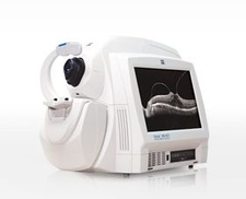 Zeiss Cirrus HD-OCT
Cirrus HD-OCT enables your optometrist to perform multiple analyses for glaucoma.
The three most common tests are:
- Retinal Nerve Fibre Layer (RNFL) Analysis reveals the thickness of the layer of the retina that contains nerve fibres that travel up the optic nerve. If glaucoma is present, that layer may gradually lose thickness.
- Macular Thickness Analysis examines the condition and thickness of the macula, which is the part of the retina that provides central vision. Thinning of the macula is a possible sign of glaucoma progression.
- Optic Nerve Head Analysis reveals the structure of the optic nerve where it originates in the retina. With glaucoma, the “cup” in the optic nerve may enlarge.
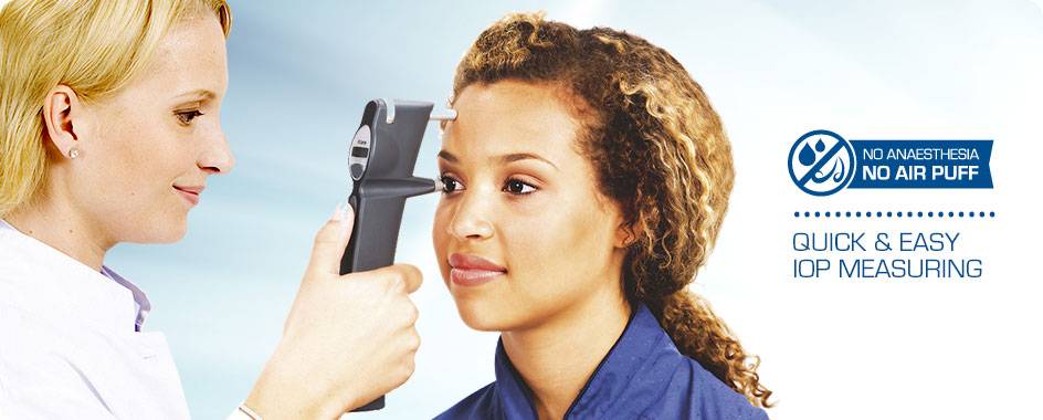
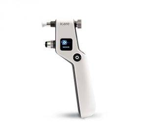 iCare Tonometer
iCare Tonometers for easy, accurate and patient-friendly intra-ocular pressure measurement.
iCare tonometers are based on unique, patented rebound technology, in which a very light and small probe is used to make a momentary contact with the cornea. No specialized skills for its use the quick and painless measurement is barely noticed by the patient and and any anesthesia or inconvenient air puffs are not needed at all.Icare technology has been proven accurate and reliable by several clinical studies which demonstrates the same accuracy and uncompromised reliability in its measurements as the Golden standard i.e. complies to ISO 8612 tonometer standard, and better results than other hand-held tonometers presently on the market.Never before has intra-ocular pressure (IOP) measuring been this easy – and still accurate.
Visual Field Testing
A visual field test measures the range of your peripheral or “side” vision to assess whether you have any blind spots (scotomas), peripheral vision loss or visual field abnormalities.